Macular Hole: Cause and Treatment.
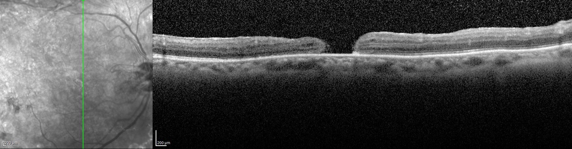
A macular hole is an opening in the central portion of the retina, the macula. This part of the eye is responsible for seeing fine detail such as letters on a page or numbers on a telephone. Macular holes occur most frequently in healthy people, most of whom are women in their 60’s and 70’s.
The inside cavity of the eye is filled with a gel called the vitreous, similar to how air fills the inside of a basketball. Macular holes occur when the portion of the vitreous that lies on the macula spontaneously contracts, pulling some of the macula with it. If the pulling is strong enough, a hole develops along with a small area of retinal detachment, causing loss of fine vision.
How is a macular hole treated?
In rare cases, a macular hole closes on its own. In severe cases (Stage 3 & 4), doctors will perform vitrectomy surgery. They remove the vitreous gel/fibrous membrane responsible for the pulling causing the macular hole. A temporary gas bubble is placed in the eye to seal the hole. The bubble is then absorbed by the body over 10 – 14 days and is replaced by saline produced naturally by the eye. The gas bubble then puts pressure on the macula, causing the hole to flatten and close. Part of the recovery involves lying face down for several days.
The technique uses a dye (indocyanine green or brilliant blue) and an instrument developed by Yasuo Tano in Japan made from micro diamond dust allows the tissue causing the macular hole to be removed. The success rates is greater than 95%.
Another condition that could lead to macular hole is Vitreomacular Traction Syndrome (VMT). Macular holes caused by VMT are treated with Ocriplasmin, a recombinant protease with activity against fibronectin and laminin, components of the vitreoretinal interface. It received FDA approval in 2012.